


Endodontics (root canal treatment)
High-quality endodontic treatment is made possible by modern devices such as RVG (radiovideography), apex locators, systems for mechanical enlargement of root canals, systems for thermal root canal filling and a laser for root canal treatment… The treatment results are shown to the patient immediately after treatment, which is made possible by digital X-ray imaging of the affected areas. All X-ray examinations are performed on our premises, which reduces the burden on patients and, above all, enables immediate diagnosis and presentation of treatment results.
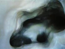
Root canal treatment
Root canal treatment comprises fundamental procedures that make it possible to preserve teeth despite advanced dental disease that has caused irreversible damage to the vital dental pulp. The causes of such damage vary, but tooth decay remains the leading cause. The disease causes severe inflammation of the pulp, which may be accompanied by characteristic severe toothache, and the pulp subsequently dies. The result is therefore dead tissue within the hard dentine-enamel chamber of the tooth, which provides an ideal breeding ground for bacteria. We say that so-called gangrene has developed.

Unfortunately, the bacteria do not remain only inside the tooth but slowly spread to the periodontal tissues as well. The condition may again be accompanied by pain and swelling of the soft periodontal tissues. Even in cases where the disease is not accompanied by acute local signs, there is a risk of bacteria and their toxins being released slowly into the bloodstream, which may result in distant signs of disease. This is referred to as the development of a chronic focus of infection. It follows from the above that, when treating such a tooth, the dead pulp must be removed and the resulting empty space filled with materials that prevent bacterial growth. This method of treatment is therefore called root canal treatment. The success of this treatment is not always complete and depends on many factors, including the precision and capabilities of the practitioner and their equipment.

In recent years, technological development has opened up new possibilities in endodontics, or root canal treatment, as well. Magnifying devices and operating microscopes give us a better view of the pulp chamber and therefore enable more successful identification of the microscopically small entrances to root canals where remnants of dead tissue are concealed. Well-known researchers report a success rate of as much as 70% in locating the most difficult-to-access root canal in upper first molars with the aid of an operating microscope, whereas without its use they were successful in just under 20% of cases. Complete removal of dead tissue is the basic condition for relieving the patient of persistent pain and preventing later problems. To provide a clearer illustration, we present the case of a forty-year-old woman who came to our dental office with pain in the region of the lower first molar (36), which had persisted since the first endodontic treatment two years earlier. She reported a tearing sensation in the region of the tooth, and the tooth was also sensitive to temperature stimuli. During the examination, we found that the tooth had a large amalgam filling that slightly overhung the gingival area, which also caused persistent bleeding during cleaning. We found no swelling or pain in the gum area.
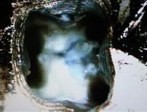
The X-ray showed adequate filling of three root canals, which is generally usual. A non-homogeneous and porous existing filling and the presence of a fourth canal, which had not been found during the first root canal treatment two years earlier, were suspected. We decided to repeat the root canal treatment. After local anaesthesia, we removed the old filling and the remnants of the previous root canal fillings. Using an injection needle, we rinsed the canals with bacteria-removing solutions (NaOCl). With the aid of magnifying devices, we discovered the entrance to the previously undetected fourth root canal. Because the canal bled during treatment with endodontic files, we concluded that living soft dental tissue was still present in the canal, which was the main reason why the first treatment had been unsuccessful. After a thorough examination of the other previously treated root canals, we found that their fillings were porous. We cleaned all the canals, rinsed them with medication and introduced an antibiotic paste into them. We temporarily sealed the tooth with glass ionomer cement and a semi-soft filling and scheduled the patient for another appointment one week later.
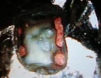
In the second phase, we repeated the procedures from the first phase, but instead of introducing antibiotics, we placed a paste with a high pH value (Ultracal) into the canals, meaning that its high alkalinity creates unfavourable conditions for bacteria in the canals. We scheduled the patient for another appointment six weeks later.
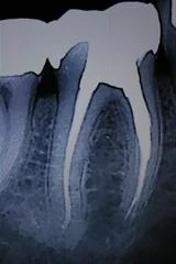
The patient arrived for her third appointment relaxed and satisfied and did not complain of pain in the tooth concerned. We reopened the tooth, removed the remnants of the medicated root canal dressing placed during the second phase and dried the root canals. Examining the root canals with the aid of magnification reduces the likelihood of leaving remnants of the dressing in the canal. We took another image of the tooth with a digital X-ray device and, with the aid of silver points inserted into the canals, precisely determined their actual length. We definitively filled the tooth with plastic materials and additionally with a filling material (AH 26), which seals the canals precisely and reduces the possibility of renewed bacterial growth. Finally, we checked the accuracy of the filling with another X-ray image, which showed a precise and homogeneous filling.
The patient arrived for her third appointment relaxed and satisfied and did not complain of pain in the tooth concerned. We reopened the tooth, removed the remnants of the medicated root canal dressing placed during the second phase and dried the root canals. Examining the root canals with the aid of magnification reduces the likelihood of leaving remnants of the dressing in the canal. We took another image of the tooth with a digital X-ray device and, with the aid of silver points inserted into the canals, precisely determined their actual length. We definitively filled the tooth with plastic materials and additionally with a filling material (AH 26), which seals the canals precisely and reduces the possibility of renewed bacterial growth. Finally, we checked the accuracy of the filling with another X-ray image, which showed a precise and homogeneous filling.
In the next phase, we restored the tooth with a radicular post and a metal-ceramic crown. Six months later, we scheduled the patient for another X-ray examination, which showed healthy bone in the region of the tooth root tips, providing final satisfaction and confirmation that the treatment had truly been successful.
With this example, we wanted to demonstrate the method, duration and complexity of root canal treatment and, above all, the importance of using magnifying devices or operating microscopes in modern dentistry. According to the professional literature, an operating microscope makes it possible to locate hidden entrances to the root canals of upper first molars in as many as 90% of cases; without its use, the practitioner would otherwise overlook them and leave dead tissue in the tooth, together with all the consequences described at the beginning of this article. All of this raises the question of the ethics of treatment without the use of every available means for treating a particular problem and, at the same time, the question of the actual cost of such complex procedures. We leave that question to those who decide how much money is allocated to each of us for the treatment of dental diseases.
With this example, we wanted to demonstrate the method, duration and complexity of root canal treatment and, above all, the importance of using magnifying devices or operating microscopes in modern dentistry. According to the professional literature, an operating microscope makes it possible to locate hidden entrances to the root canals of upper first molars in as many as 90% of cases; without its use, the practitioner would otherwise overlook them and leave dead tissue in the tooth, together with all the consequences described at the beginning of this article. All of this raises the question of the ethics of treatment without the use of every available means for treating a particular problem and, at the same time, the question of the actual cost of such complex procedures. We leave that question to those who decide how much money is allocated to each of us for the treatment of dental diseases.
✦ This content was translated from the original with the assistance of an AI translation system.