


Modern Dental Radiology
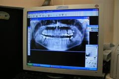
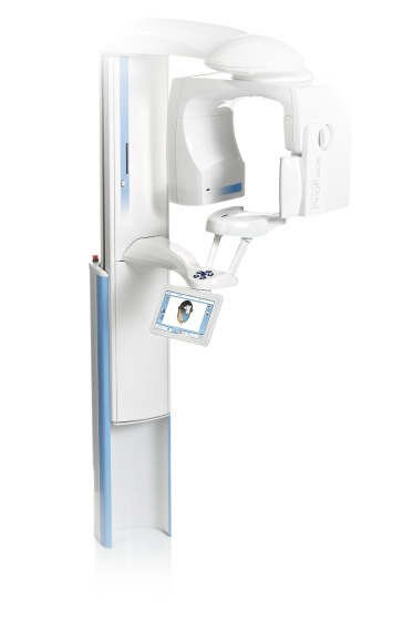
The use of X-ray images is essential for successful treatment and prevention. We distinguish between local dental X-rays and panoramic images, or orthopantomograms. The use of digital radiology has reduced the burden on patients and practitioners, as radiation doses are considerably lower compared with conventional radiology. Digital technology does not require developing chemicals, which is also good news for environmental protection.
Modern dental medicine today is unimaginable without modern X-ray imaging of the teeth and periodontal tissues. In the past, conventional X-ray machines were used for this purpose, in which the X-ray sensor was conventional mercury film, similar to that used in a camera. Although such images are very precise, developing the film takes a great deal of time, while the chemical reactions involved are highly harmful to people and the environment. The harmful effects of imaging on the person exposed are relatively high compared with modern digital radiology, which uses a digital sensor instead of a conventional sensor and is much more sensitive to X-rays. The harmful effects on the person exposed can therefore be much lower, conventional image development is unnecessary, and the image appears on the LCD monitor within a few seconds. Patients can have the image explained to them, as the monitor is usually mounted directly on the dental chair. Root canal treatment is therefore easier for them to accept, as they can also participate to some extent in treatment decisions.
Modern dental medicine today is unimaginable without modern X-ray imaging of the teeth and periodontal tissues. In the past, conventional X-ray machines were used for this purpose, in which the X-ray sensor was conventional mercury film, similar to that used in a camera. Although such images are very precise, developing the film takes a great deal of time, while the chemical reactions involved are highly harmful to people and the environment. The harmful effects of imaging on the person exposed are relatively high compared with modern digital radiology, which uses a digital sensor instead of a conventional sensor and is much more sensitive to X-rays. The harmful effects on the person exposed can therefore be much lower, conventional image development is unnecessary, and the image appears on the LCD monitor within a few seconds. Patients can have the image explained to them, as the monitor is usually mounted directly on the dental chair. Root canal treatment is therefore easier for them to accept, as they can also participate to some extent in treatment decisions.
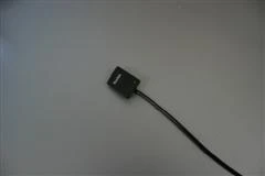
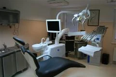
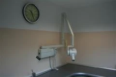
Digital radiography is thus successfully replacing conventional radiography, as practitioners have begun to recognise the advantages of its use. It has an exceptionally high resolution of 15 lp/mm to 20 lp/mm, as the pixel size is only 19.5 microns. Various sizes of square-shaped sensors are available and are easy to insert into the oral cavity. Images can easily be stored on disks and quickly retrieved from data storage. Compared with conventional radiology, storage requires extremely little space. Practitioners can exchange images with one another over the internet.
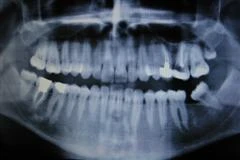
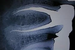
The software supporting this type of imaging enables precise relative measurements, which are crucial in diagnostic and therapeutic procedures. It has thus been established that programs based on determining the optical density of the imaged material successfully identify minimal differences in the mineralisation of hard dental tissues and detect early carious processes in the interdental spaces, where they are most commonly missed during clinical examinations. Magnification of digital X-ray images enables precise endodontic or root canal treatment, as it makes it easier, or even possible, to assess key areas that are usually hidden from view. To use X-ray equipment, every practitioner must obtain the appropriate permits issued by the Slovenian Radiation Protection Administration. The following are required for a permit: an equipment inspection report, a medical certificate, regular dosimetry and a radiation protection course. Patients can verify all the documentation obtained with the practitioner. Since there is a misconception among patients that dental X-ray imaging is highly harmful, the following data show the average doses—and therefore the level of harmful exposure—for comparison:
- Panoramic dental image: 0.011 mSv
- Intraoral dental image: 0.010 mSv
- Pelvic CT: 10 mSv
- Chest X-ray: 0.04 mSv.
It follows from the above that the amount and importance of the information obtained from individual dental imaging procedures—which are, of course, professionally justified—are much greater than the negative effects of the image. The balance shifts even further in favour of imaging when a digital X-ray machine is used, as the harmful effects of radiation are approximately ten times lower than with conventional imaging. Modern X-ray imaging thus places a considerably lower burden on the patient and practitioner and, because toxic chemicals are not used, also on the environment.
Sources for further reading:
- Farman AG, Rarman TT. RVG-ui: a sensor to rival direct exposure intra oral X-ray film. Int J Comput Dent. 1999;2:183-96.
- Mouyen F et al. Presentation and physical evaluation of RVG. Oral Surg Oral Med Oral Pathol. 1998; 68: 238-42.
- Forner L et al.Digital radiology and image analysis for approximal caries diagnosis. Oper Dent. 1999;24:312-5.
- Dagenais ME, Clark BG. Receiver operating charasteristics of RVG. Oral Surg Oral Med Oral Pathol Oral Radiol Endod. 1995;79:238-45.
- Wenzel A et al. Accuracy of caries diagnosis in digital images from chandarged coupled device and storage phosphor systems: an in vitro study. Dentomaxillofac Radiol. 1995 ;24:250-4.
- Hayakava et al. Radiation dosage reduction in general dental practise using digital intraoral radiographic system. Bull Tokyo Dent Coll. 1997; 38:21-5.
- Pfeiffer et al. Effects of different exposure values on diagnostic accuracy of digital images. Quintesence Int. 2000; 31:257-60.
- Ledy BG et al. Interpretation of endodontics file lenghts using RVG. J Endod. 1994; 20: 542-5.
- Scarfe WC, Fana CR Jr, Farman AG. Radiographic detection of accessory/lateral canals: use of RVG and Hypaque. J Endod. 1995;21:185-90.
- Yokota ET et al. Interpretation of periapical lesions using RVG. J Endod. 1994; 20: 490-4

✦ This content was translated from the original with the assistance of an AI translation system.